Amadeus C. S. de Alcântara
School of Mechanical Engineering and Center for Computing in Engineering & Sciences – University of Campinas
![]()
How do bones fracture? What influence does osteoporosis have on bone fracture? How to identify the risk of bone fracture and anticipate osteoporosis related fractures? These are questions which CCES’s researchers have been trying to answer with the use of computer simulations. Estimation of bone fracture risk is still an open field of research that needs further development. Bone tissue is a very complex material not yet completely understood.
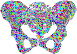
Pelvis computational model created from computer tomography images.
For those reasons, some may ask – why study bone fracture? Simply because due to accelerated development in technology and medicine, people are living longer worldwide, and yet, a longer life does not necessarily mean a healthier life. The rising average life expectancy implies that a greater fraction of the population will be afflicted by osteoporosis, a chronic condition which decreases bone toughness, increases fracture risk, and affects mainly the elderly population. If current medical diagnosis technologies do not improve, osteoporosis-induced bone fracture will become a heavier social and financial burden for health care systems globally.
By understanding how bone fails and by correlating this understanding to early signs of tissue deterioration, bone fracture initiation may be identified before its usual clinical diagnosis. Medical imaging techniques, especially computer tomography, are able to provide computational models of affected bones, displaying accurate geometry and material properties of each individual patient.
Bone fracture is a phenomenon that initiates at the molecular scale. The molecular structure of a fragile bone, when hit by a strong impact load, starts to display failures. When several (millions) atom bondings start to break, fracture propagates and becomes visible. Try to think of a crack, a small hole, in the bone, but at the microscale. If this crack propagates up to the macroscale, the bone will break.
Simulating a crack propagation starting at the molecular scale and developing up to the macroscale is a challenging process. When simulating a bone fracture, it is important to take into account the smallest structures of the bone and their relationship with largest structures. A proper representation of the bone starting from the atoms, passing through the molecular structures, and up to the whole macroscopic bone is fundamental. This is called multiscale modelling.
Once built, the multiscale model uses computational methods to calculate traction and displacement at each pre-selected point of the structure and to foresee the possibility of failure at all length scales.
Molecular models are used to simulate the motion and behaviour of atoms and molecules, where all processes begin. Numerical methods help us estimate the possibility of a fracture. This kind of multiscale bone fracture modelling requires advanced calculations and high computational power.
The group directed by Prof. Paulo Sollero (School of Mechanical Engineering – UNICAMP), together with the groups coordinated by Prof. Munir Skaf (Institute of Chemistry – UNICAMP) and Prof. Lucia Costa Paiva (School of Medical Sciences – UNICAMP) simulates fracture in osteoporotic bone models created from computed tomography taken directly from the patients. The groups seek, using the boundary element method and performing molecular dynamics simulations, to better understand how and when bones, especially osteoporotic bones, start to fail. The ultimate goal is to create a computational tool based on mathematical methods capable of assisting healthcare personnel to estimate the bone fracture risk of a patient, with particular emphasis on those afflicted by osteoporosis.
Associated Scientific Research:
A. C. S. Alcantara, “Implementierung verschiedener Algorithmen zur automatisierten Berechnung und Zuweisung von Materialgesetzen von CT-Daten auf FE-Netze”, 2017.
D. M. Prada, A. F. Galvis, A. C. Alcântara and P. Sollero, “3D Boundary element meshing for multiscale bone anisotropic analysis”, European Journal of Computational Mechanics, 2018.
G. Osterhoff, E. F. Morgan, S. J. Shefelbine, L. Karim, L. M. McNamara, and P. Augat, “Bone mechanical properties and changes with osteoporosis”, Injury, 2016.